Not All Plaque is Equal
- || Tags: Arterial Plaque, CLEERLY, heart attack, heart health, Prevention

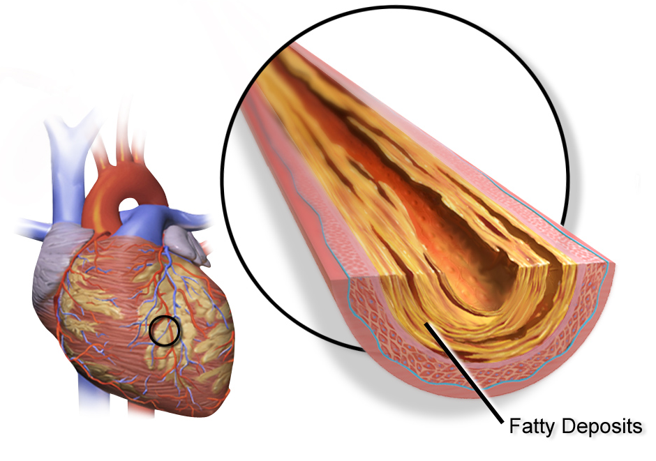
Heart attacks are principally caused by artery plaque becoming dislodged, causing an arterial obstruction, and a significant number of patients who suffer a heart attack never have any warning signs.
The majority of cardiac emergency events can be traced back to noncalcified plaque, a buildup of soft deposits embedded deep within the walls of the heart’s arteries, undetectable by angiography or cardiac stress tests — and prone to rupture without warning, according to the CDC.
Determining what type of plaque built up in your arteries is taking place can be a significant step in helping prevent a potentially fatal cardiac event; and the safest, most non-invasive method for determination is through a CT or MRI scan of the heart.
“The importance of quantifying plaque is critical because total plaque burden is considered the most important predictor of coronary events,” Melvin Clouse, M.D., Ph.D., Emeritus Chairman of the Department of Radiology and Director of Radiology Research at Beth Israel Deaconess Medical Center in Boston, and Deaconess Professor of Radiology at Harvard Medical School, writes in a study about plaque build-up and detection. “Furthermore, the rupture of soft noncalcified plaque has been implicated as the cause of heart attack.”
Dr. Clouse, an interventional radiologist and researcher, has authored several studies on plaque phenotypes and ways to determine the risk of heart attack, as well as ways to mitigate such risk.
Exercise stress testing and coronary angiography, the standard methods for diagnosing atherosclerosis and heart attack risk, both work by visualizing the lumen, the channel through which blood flows.
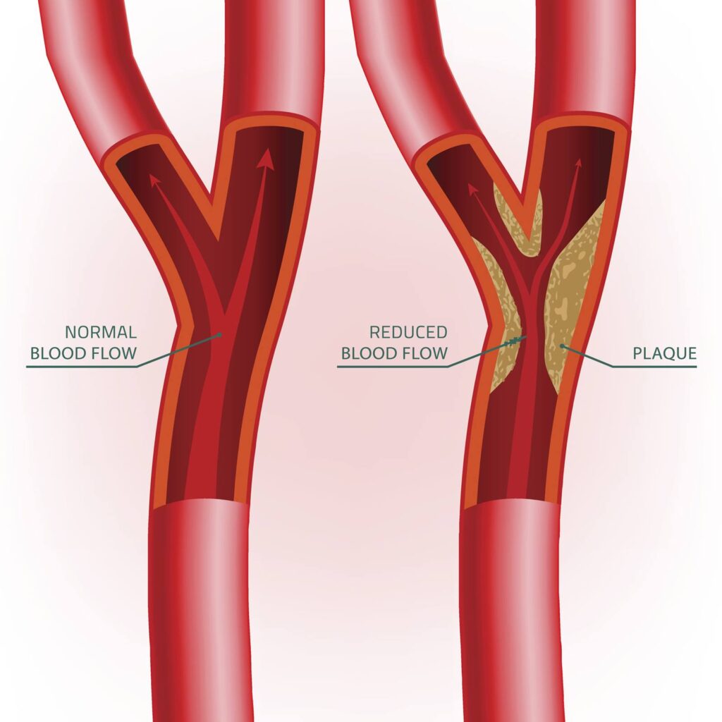
However, because the lumen also increases in size as plaque progresses, coronary artery disease may go undetected until late in the disease process. And, Clouse writes, “Because soft plaque buildup may not significantly narrow the lumen, conventional angiography and stress tests fail to provide a complete picture of plaque accumulation.”
Now, technology exists allowing patients to have their heart scanned to both measure the amount of plaque build-up, determine what type of plaque exists, and in concert with their cardiologist or internist, prevent a heart attack or stroke before it happens.
“[S]canners have made it possible to detect noncalcified plaque,” Clouse writes. “However, … accurate and reproducible measurements of this plaque was difficult and time-consuming [before] a new technique that would overcome these obstacles [was finally developed].”
Ascend Imaging Center, in partnership with Cleerly, a laboratory in Colorado that has pioneered the use of artificial intelligence in MRI heart scanning, can now provide you and your physician a detailed analysis of plaque amount and phenotype, significantly changing the way preventative medicine is practiced.
For more information on the Cleerly Heart Scan and other preventative analysis tests, contact Ascend Imaging today at (248) 595-8404.
Conveniently located at the northwest corner of Telegraph and 12 Mile Roads, inside the Comerica office tower, Ascend Imaging Center is 20 minutes from nearly anywhere in the metropolitan Detroit area — and your first stop for heart health.


